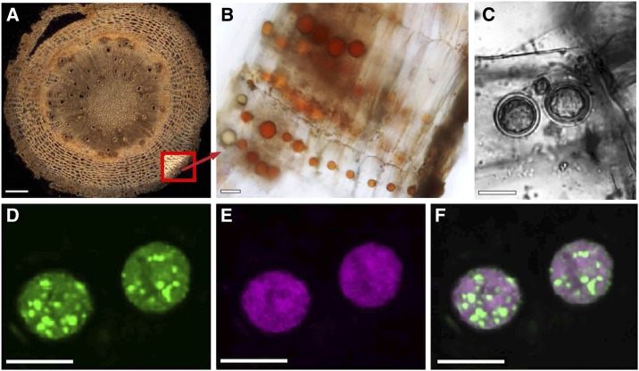
Figure 1.
Localization of oil bodies within the root cork of C. forskohlii. A, Cross section of entire root with thick-fissured cork. Bottom right inset, the location of cork cells. B, Rows of cork cells each with one prominent oil body. C to F, Confocal imaging of Nile Red-labeled oil bodies. C, Transmitted light image of a cork cell with two oil bodies. Fluorescence images of the same oil bodies showing discrimination between neutral lipids (green fluorescence, D) and polar lipids (magenta fluorescence, E). F, Overlay of the two fluorescence images. Bars = 200 µm (A) and 10 µm (B–F).

