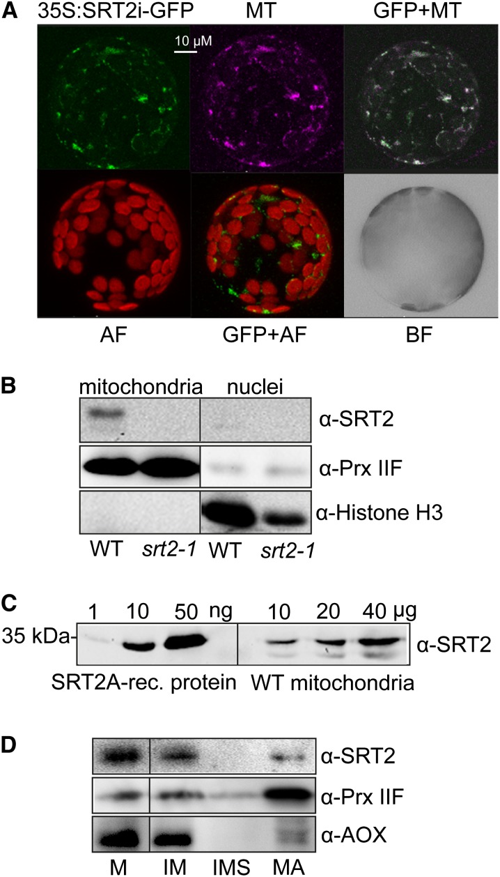
Figure 2.
SRT2-encoded proteins localize to mitochondria in Arabidopsis. A, Arabidopsis protoplasts showing GFP localizations (green) for a full-length SRT2 (plus introns)-GFP fusion construct (35S:SRT2i-GFP). Protoplasts were isolated from stable Arabidopsis transformants. Purple indicates the mitochondrial localization of MitoTracker (MT). GFP+MT indicates overlay image of 35S:SRT2i-GFP and MitoTracker; AF indicates autofluorescence of chloroplasts; and BF indicates bright-field image of protoplast. B, Western-blot analysis of SRT2 protein in mitochondrial and nuclear fractions. Antisera against PRX IIF was used as mitochondrial marker and histone H3 as nuclear marker, respectively. C, Western-blot analysis of SRT2 proteins in isolated mitochondria. Protein extracts of isolated mitochondria from wild-type (10, 20, and 40 µg) seedlings as well as recombinant 6× His-SRT2A protein (1, 10, and 50 ng) were analyzed by western blotting using SRT2 antiserum. D, Western-blot analysis of SRT2 protein in subfractionated Arabidopsis wild-type mitochondria. Ten micrograms of protein were loaded for each fraction. Antisera against Alternative Oxidase1 and PRX IIF were used as controls for the inner membrane and matrix proteins, respectively. M, Mitochondria; IM, inner mitochondrial membrane; IMS, intermembrane space; MA, matrix.

