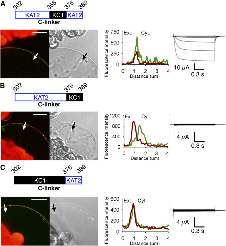
Figure 2.
The extreme C-terminal part of the C-linker plays a role in KAT2 surface expression. Three different channel chimeras (A–C) were obtained by sequence swapping between KAT2 and AtKC1. The regions from KAT2 and AtKC1 are depicted in blue and black, respectively. Surface expression and activity at the PM of each chimeric channel were investigated by subcellular localization of GFP fusions in tobacco protoplasts and current recordings in X. laevis oocytes as described in the legend to Figure 1. A white arrow on the confocal image marks the position of the analyzed section crossing the PM and pockets of cytoplasm. Bars = 10 μm.

