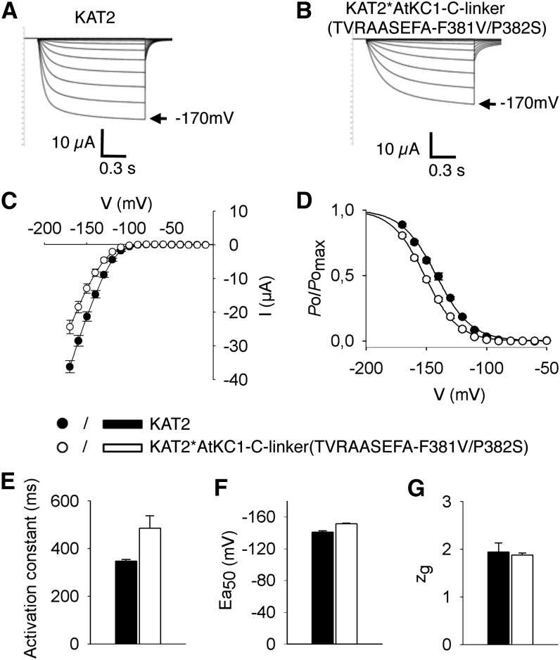
Figure 7.
Electrophysiological characterization of the KAT2*AtKC1-C-linker(TVRAASEFA-F381V/P382S) chimera in X. laevis oocytes reveals KAT2-like functional properties. Oocytes were injected with 15 ng of either KAT2 or KAT2*AtKC1-C-linker(TVRAASEFA-F381V/P382S) cRNA. Currents were recorded in 100 mm K+ external solution. A and B, Representative current traces in oocytes expressing KAT2 (A) or KAT2*AtKC1-C-linker(TVRAASEFA-F381V/P382S) (B). C to G, Steady-state current (I)/voltage (V) curves (C), relative open probabilities (D), activation time constants (E), half-activation potentials (Ea50; F), and equivalent gating charge (zg; G) of channel activity in oocytes expressing KAT2 (black bars, black circles) or the KAT2*AtKC1-C-linker(TVRAASEFA-F381V/P382S) chimera (white bars, white circles). Means ± se (n = 10–15) were obtained using the same oocyte batch. Applied activation membrane voltages ranged from +0 to −170 mV (increments of 10 mV; holding potential, 0 mV; deactivation potential, −60 mV).

