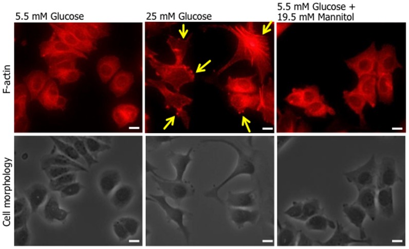
Figure 2. The formation of lamellipodia and morphology in MCF-7 cells cultured with high glucose level.
MCF-7 cells plated on Collargen IV-coated coverslips were cultured with normal (5.5 mM), high (25 mM) D-glucose concentration, or an osmotic control (5.5 mM D-glucose plus 19.5 mM D-mannitol) medium for 24 h. Cells were fixed with 3.7% formaldehyde, permeabilised with 0.1% Triton X-100 and stained with Alexa Fluor®546-labelled phalloidin. Upper panels; fluorescence images, lower panels; phase-contrast images. Representative results of 3 independent experiments are shown. Arrows show representative lamellypodia formations. Bars: 10 µm.

