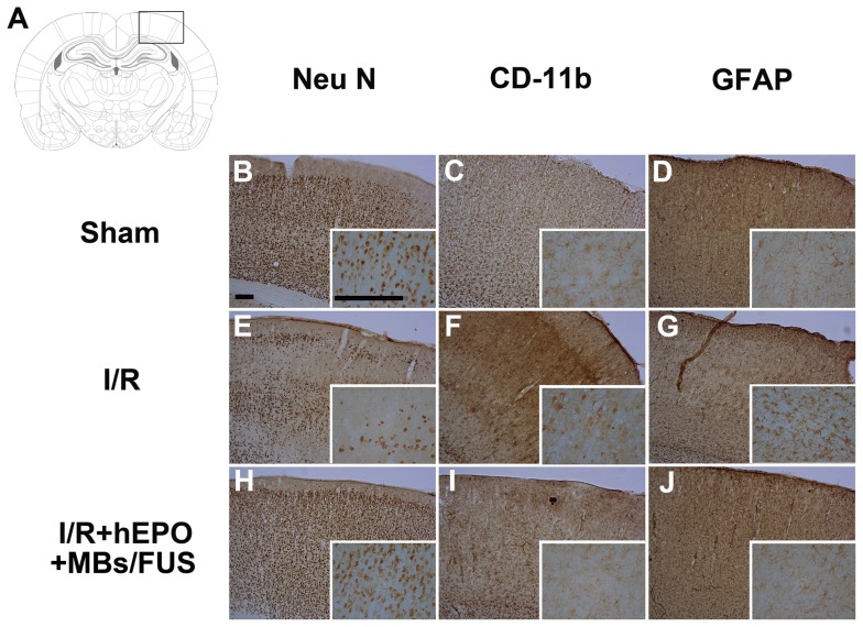
Figure 4. hEPO+MBs/FUS inhibits the ischemia/reperfusion-induced neuronal death and inflammation in rat experiments.
Immunohistochemical staining of NeuN, CD-11b, and GFAP was performed 24 h after I/R. (A) illustrated the position of FUS sonication. In NeuN staining (B, E, and H), the I/R group showed a marked loss of neurons, whereas, neurons were intact in the sham and I/R+hEPO+MBs/FUS groups. In CD-11b staining (C, F, and I), the I/R group showed microglia activation (condensed nuclei), whereas the sham and I/R+hEPO+MBs/FUS groups showed ramified microglia. In GFAP staining (D, G, and J), there was an increase of GFAP in the I/R group but not in the sham and I/R+hEPO+MBs/FUS groups. (Scale bar = 200 µm).

