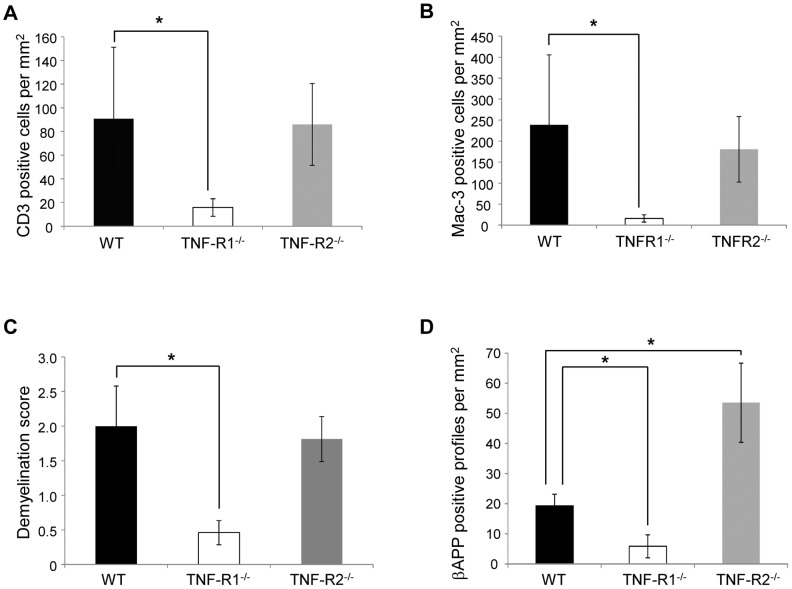
Figure 2. Histopathological analysis confirmed that EAE was less severe in TNFR1-/- mice in comparison to both WT and TNFR2-/- mice.
Spinal cords were analysed at day 21 of EAE for signs of inflammatory infiltration, demyelination and axonal degeneration. TNFR1-/- mice had significantly less infiltration of both CD3-positive T cells (A) and Mac-3-positive activated microglia/macrophages (B) when compared to WT and TNFR2-/- mice. Furthermore, TNFR1-/- mice also had significantly less demyelination, as assessed by LFB staining (C) and axonal damage, as assessed by accumulation of β-APP-positive axonal profiles (D), when compared to both WT and TNFR2-/- mice. Conversely, TNFR2-/- had significantly increased numbers of β-APP positive damaged axons compared to WT mice.

