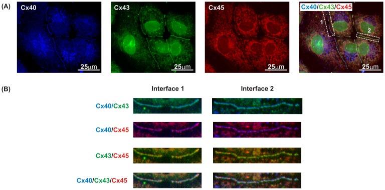
Figure 4. Co-localisation of Cx40, Cx43 and Cx45 in clone 6.
(A) Each connexin can be seen in the individual channels within the same area and the triple labelling image was generated by superimposing the image for each connexin subtype. (B) To demonstrate the varying degrees of co-localisation at each cell interface, sections 1 and 2 highlighted from triple labelling were separated into dual images of Cx40/Cx43, Cx40/Cx45, Cx43/Cx45 and triple image of Cx40/Cx43/Cx45 as indicated on the left panel of the image.

