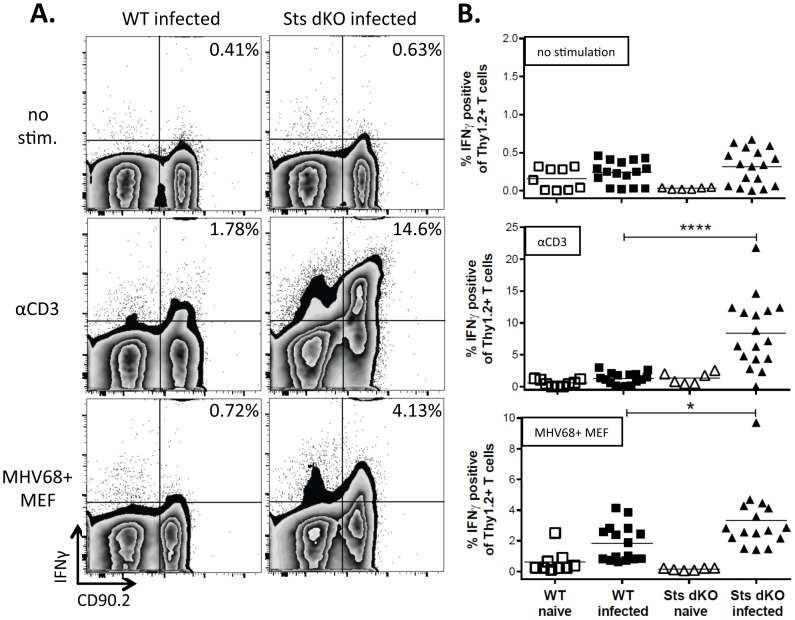
Figure 1. Increased IFNγ response to infected cells in the absence of Sts1 and Sts2.
Sts dKO and C57BL/6 WT mice were infected 1000 PFU of MHV68 by the intranasal route and spleens were harvested 28 dpi. Splenocytes were left untreated or stimulated with 1 ug/ml αCD3 antibody or cocultured with gamma-irradiated MHV68-infected MEFs, both overnight in the presence of monensin. Cells were stained with the T cell marker CD90.2 and the intracellular cytokine IFNγ and analyzed by flow cytometry. (A) Representative flow plots of IFNγ expression in cells stained for CD90.2 are shown for each genotype and culture condition. (B) Scatter plot summary of the percentage of T cells producing IFNγ+ after overnight stimulation. Symbols represent data from individual mice. * = p<.05, **** = p<.0001.

