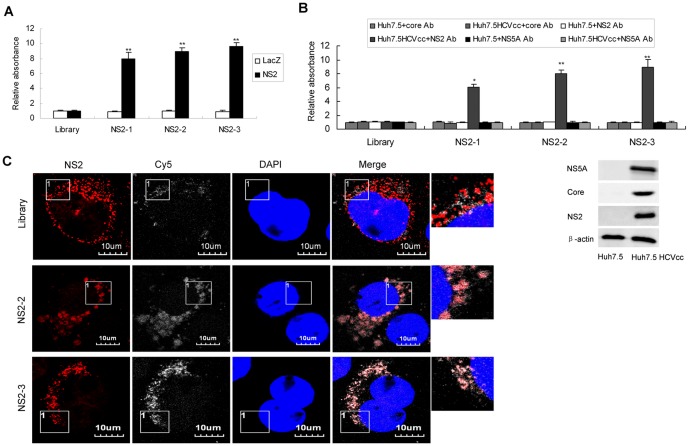
Figure 2. Binding affinity of selected aptamers against NS2.
(A) The binding affinity of aptamers NS2-1, NS2-2 or NS2-3 to recombinant NS2 protein. Biotin-labeled each aptamer was added to the microtiter plate previously coated with streptavidin. Purified NS2 protein or control LacZ protein was added to the plates. ELONA assay was performed. The absorbance was normalized to the library and represented means of 3 different experiments performed in triplicate. ** P<0.01 verse library. (B) The binding affinity of aptamers against NS2 to NS2 protein in the lysates of HCV-infected Huh7.5 cells. The plates were treated as described in part A. Lysates of HCV-infected Huh7.5 cells or control Huh7.5 cells were added to the plates. Mouse anti-NS2, anti-core or anti-NS5A monoclonal antibody was used. The absorbance was normalized to the library and represented means of three independent experiments performed in triplicate (upper). * P<0.05, ** P<0.01 verse library. Core, NS2 or NS5A protein in the lysates of HCV-infected Huh7.5 cells and naïve Huh7.5 cells was detected by western blot using antibodies against HCV core, NS2 or NS5A respectively (lower). (C) Co-localization of NS2-specific aptamers with NS2 protein in the HCV-infected hepatocytes. HCV-infected Huh7.5 cells were treated with Cy5-labeled aptamers for 24 hours. The cells were fixed with ice-cold acetone for 10 minutes at −20°C. The cells were washed with PBS, blocked with 1:50 goat serum for 30 minutes at room temperature and then incubated for 1 hour with mouse monoclonal anti-NS2 antibody. The cells were stained with Texas Red-labeled goat anti-mouse for 45 minutes respectively at room temperature. The nuclei were counterstained with DAPI. Fluorescent images were obtained under fluorescent microscope. Identical setting was maintained for images capture. Representative images are shown.

