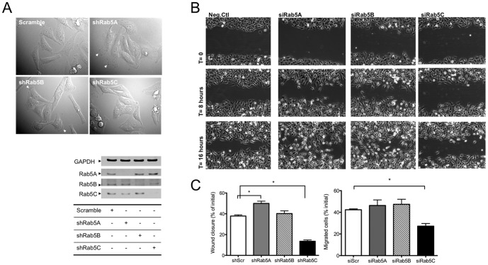
Figure 1. Rab5C depletion significantly inhibits cell migration.
A) DIC images of stable Rab5 isoform knock-down (KD) HeLa cells taken with light microscope at 40X magnification (left panel). Arrows indicate membrane ruffles. KD of Rab5 isoforms (right panel) in these stable cell lines is shown in the immunoblots following SDS-PAGE as described in Experimental Procedures. B) 0.5–1 mm width wounds were made on a monolayer of HeLa stable control or Rab5 isoform KD cells. 5–7 wounded spots in each dish were imaged with time-lapse microscope every 5 minutes for 20 hours. C) The percentage of wound closure (left panel) was calculated from images acquired at time 0 and 16 hours with ImageJ. For each sample, at least 5 images were used to calculate the percentage of wound closure in each experiment. The graph represents the Mean± S.E. from four independent experiments. U937 cells (right panel) transiently transfected with siRNA against Rab5 isoforms were seeded in the upper chamber of the Transwell plates and allowed to migrate towards 10% FBS in the bottom chamber for 24 hours. Migrated cells were measured as indicated in Material and Methods. Data are normalized to initial seeding cell numbers. The graph represents the Mean± S.E. from four independent experiments. Analysis was carried out with a one-way ANOVA, Dunnett’s post-test.(*P<0.05, ***P<0.001)

