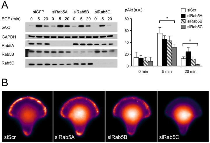
Figure 3. PI3K signaling in response to Rab5 isoform depletion.
A) HeLa cells were transfected with GFP (as negative control) or Rab5 isoform-specific siRNA. 48 hours post-transfection, cells were starved and then stimulated with EGF for indicated times. Cell lysates were subjected to SDS-PAGE and probed with antibodies as indicated. Band intensity was quantified with AlphaEaseFc 4.0 software. Bars represent the mean value ± S.E. from four independent experiments. Analysis was carried out with a two-way ANOVA, Bonferroni’s post-test. P<0.05. B) HeLa cells were transfected with indicated siRNA. 48 hours post-transfection, cells were seeded onto micropatterned coverslips coated with fibronectin, and then allowed to spread out for 2 hours in starvation medium. Starved cells were stimulated with 10 % FCS for 3 minutes and then fixed for PIP3-FITC antibody immuno-staining. Images shown here are average projections of PIP3 staining from 30–35 cells.

