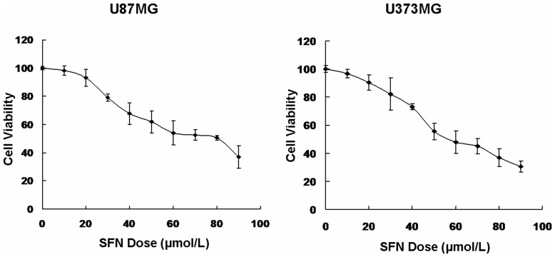
Figure 1. SFN inhibited cell viability.
An in(0, 10, 20, 30, 40, 50, 60, 70, 80 and 90 µM) for 24 h. The viability of the SFN-treated cells was measured using the MTS assay. Results were expressed as a percentage of control, which was considered as 100%. Data were reported as mean ± SD and at least three separate experiments were performed.

