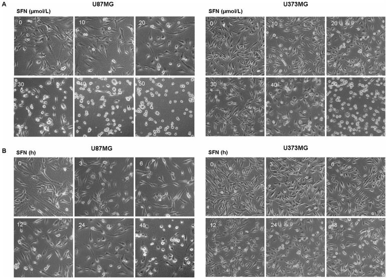
Figure 2. SFN changed cell morphology.
(A) Cellular morphological changes in U87MG and U373MG cell lines were done in a dose-dependent manner after treated with SFN for 24 h when observed by a Leica DMIRB Microscope at ×100 magnification. (B) Cellular morphological changes in U87MG and U373MG cell lines were done in a time-dependent manner after treated with 20 µM SFN when observed by a Leica DMIRB Microscope at ×100 magnification.

