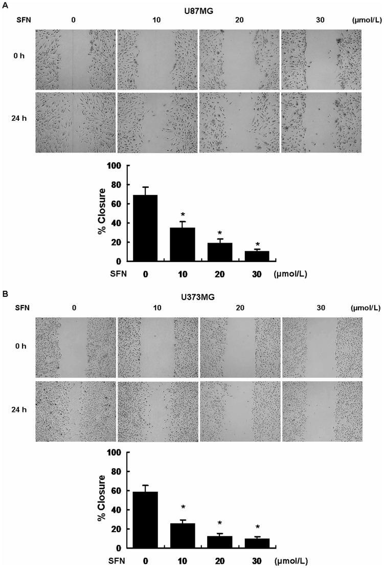
Figure 3. SFN inhibited migration in U87MG and U373MG cell lines.
Confluent U87MG and U373MG cells were scratched and incubated at different concentrations of SFN (µM). The area covered by migrating cells was recorded by phase-contrast microscopy connected to a digital camera at time 0 and 24 h. The wound closure area was calculated by measuring the diminution of the wound bed surface upon time using Image J software. Representative pictures of three independent experiments were shown. *, indicates P<0.05 versus no SFN group.

