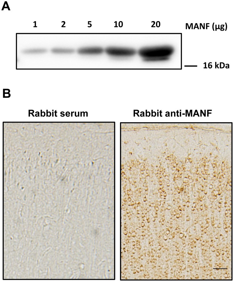
Figure 1. Detection of MANF.
A: Different amount of human recombinant MANF (1–20 µg) were loaded on a SDS-polyacrylamide gel and subjected to immunoblotting analysis using a rabbit anti-MANF antibody. B: Rat pups of PD15 were sacrificed and perfused. The brain was dissected, sectioned and subjected to MANF immunohistochemistry (IHC) using a rabbit anti-MANF antibody (1∶6,000) as described in the Material and Methods. Rabbit serum (1∶6,000) was used as a control. Images were taken from the cerebral cortex. Scale bar = 100 µm.

