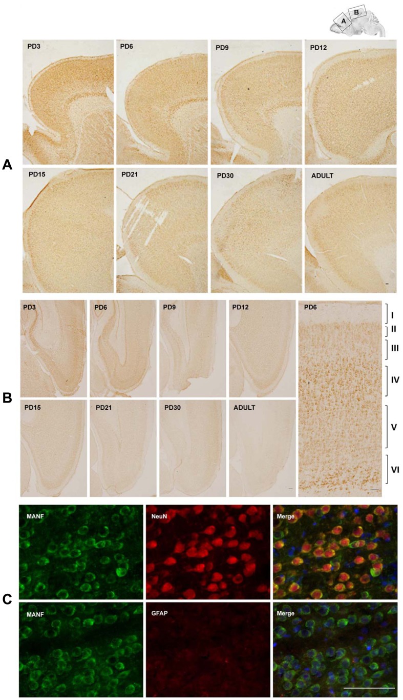
Figure 3. Study of developmental expression of MANF in the cerebral cortex by immunohistochemistry (IHC).
A and B: MANF expression in the frontal, parietal and occipital cortex. IHC images were taken from indicated cortical areas shown above. A image of higher magnification showed layers in the cortical cortex (B). C: Double labeling immunofluorescent staining was performed to determine the localization of MANF (green). Neurons and astrocytes were identified by NeuN and GFAP (red), respectively. Images were generated from layer IV of the cerebral cortex of PD15 pups. Scale bar = 100 µm.

