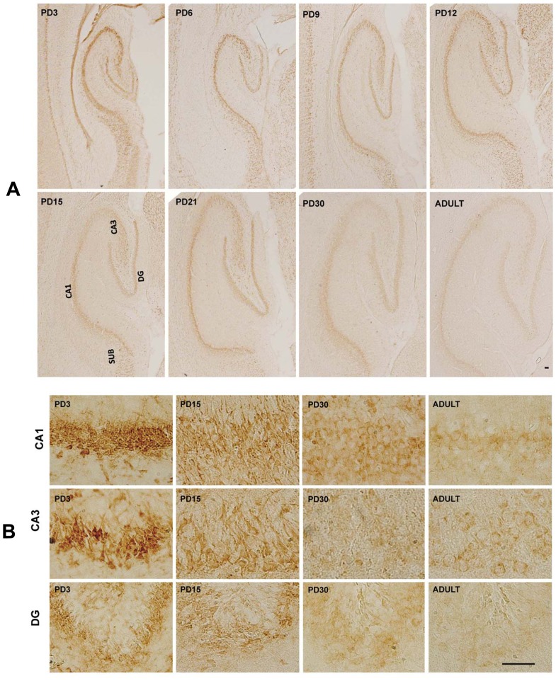
Figure 5. MANF expression in the hippocampus.
A: Rat pups were sacrificed at the indicated postnatal days. MANF expression in the hippocampus was determined by IHC. CA1 and CA3, Cornu Ammonis 1 and 3 region of hippocampus; DG, dentate gyrus; SUB, subiculum. B: Image of higher magnification showed CA1, CA3 and DG of hippocampus. Scale bar = 100 µm.

