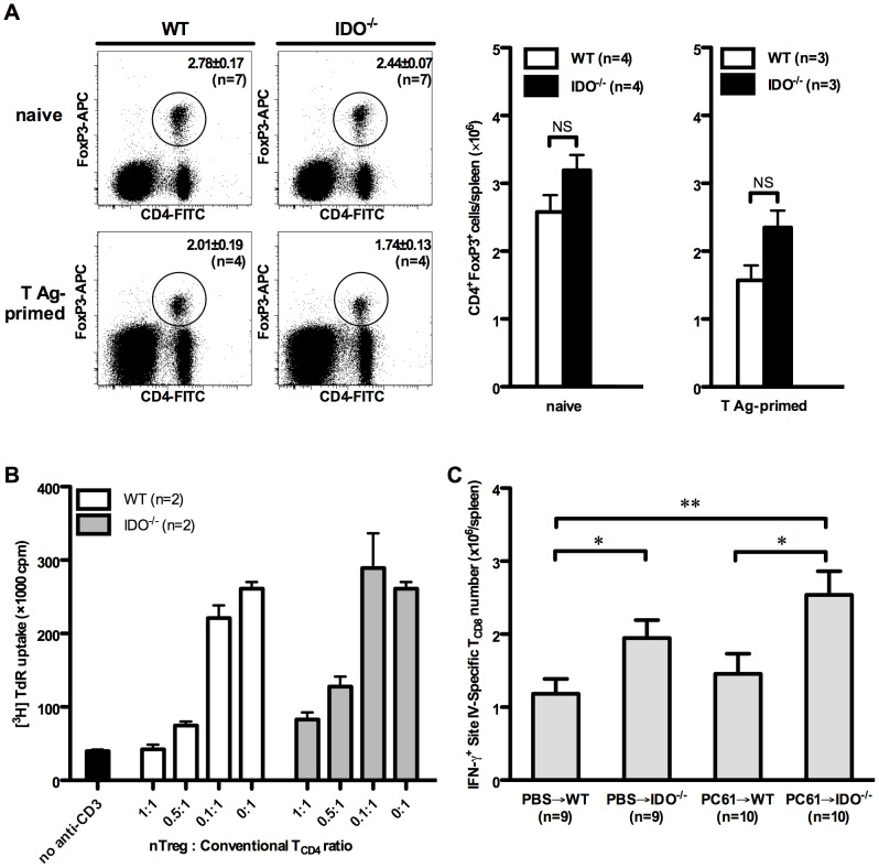
Figure 5. nTreg cells do not mediate the suppressive effect of IDO on the site IV-specific response.
(A) Splenocytes from indicated numbers of naïve WT and IDO−/− mice were stained for surface CD4 and intracellular FoxP3. In separate experiments, WT and IDO−/− mice were inoculated with C57SV cells followed, 9 days later, by cytofluorimetric determination of their splenic nTreg cell frequencies, which were used to calculate the absolute number of nTreg cells within each spleen. Representative FACS plots are shown in addition to mean nTreg cell frequencies and absolute numbers ± SEM for each group. (B) WT CD4+CD25− conventional T cells were co-cultured with γ-irradiated bone marrow-derived DCs and stimulated with an anti-CD3 mAb in the presence of varying numbers of CD4+CD25+ nTreg cells magnetically purified from WT and IDO−/− mice. T cell proliferation was measured by tritiated thymidine incorporation after 72 hours. (C) WT and IDO−/− mice were injected with an anti-CD25 mAb (clone PC61) or PBS 3 days before they were inoculated with C57SV cells. Nine days later, site IV-specific TCD8 were enumerated by ICS for IFN-γ. Values are presented as mean ± SEM for indicated numbers of mice per group pooled from 3 independent experiments.

