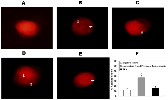
Figure 6. BPA-induced release of proteins and the initiation of apoptosis in vitro.
After liver nuclei were incubated with the supernatant from the mitochondria treated with BPA, negative control, positive control or BPA, nuclear morphologic features were assessed. (A) Normal morphologic features; (B) Positive control; (C–E) Various stages of apoptosis in liver nuclei after incubation with BPA-treated mitochondrial supernatant, showing condensation or fragmentation of chromatin in nuclei (arrow); Representative images of three separate experiments are shown (magnification, 400×). (F) Changes of apoptotic nuclei. The percentage of apoptotic nuclei was counted in five different fields of view. The results are expressed as means±S.E.M. The experiment was repeated three times. *P<0.05 compared with the negative control.

