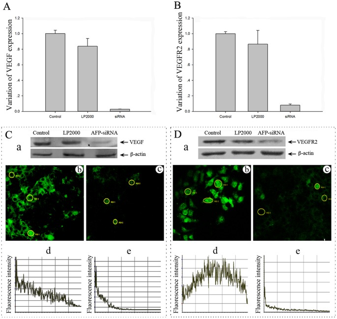
Figure 3. Effect of AFP silencing on the expression of VEGF and VEGFR-2 (KDR) in Huh-7 cells.
Cells were transfected with control siRNA, vehicle control (LP2000) and AFP-siRNA for 24, 48, and 72 hours, and the expression of VEGF (A, C, a) and VEGFR-2 (B, D, a) was examined by Real-Time PCR and Western blot. Fluorescent staining of VEGF (Cb, Cc) and VEGFR-2 (Db, Dc) following transfection with control siRNA (Cb, Db) or AFP-siRNA (Cc, Dc) are shown. Quantitative analysis of VEGF (Cd, Ce) and VEGFR-2 (Dd, De) are shown as the mean fluorescent intensity.

