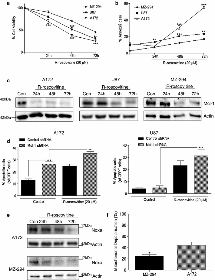
Fig. 2.

Knockdown of Mcl-1 augments R-roscovitine-induced apoptosis in A172 and U87 GBM cell lines. The GBM cells lines A172, U87 and MZ-294 were treated with R-roscovitine (20 μM) for 24, 48 and 72 h. a Cell viability was assessed using MTT assay and b the induction of apoptosis was determined by Annexin staining using flow cytometry. Data are expressed as mean ± SEM, **p < 0.01; ***p < 0.001 versus MZ-294 cells at the same timepoint. Data are from three independent experiments. c Western blot analysis of Mcl-1 expression was carried out on A172, U87 and MZ-294 cells treated with R-roscovitine (20 μM) for 24, 48 and 72 h. Actin was used as a loading control. d A172 and U87 cells were transfected with GFP-expressing Mcl-1 knockdown shRNA and subsequently treated with R-roscovitine (20 μM) for 48 h. The percentage of apoptotic GFP+ cells was determined by Hoechst staining. Data are expressed as mean ± SEM, **p < 0.01; ***p < 0.001; data are from three independent experiments. e A172 and MZ-294 cells were treated with R-roscovitine (20 μM) for 24, 48 and 72 h and the expression of Noxa was determined by western blot analysis. Actin was used as a loading control. f Mitochondrial depolarisation, indicated by the ratiometric fluorescent dye JC1, was measured in A172 and MZ-294 cells treated with Noxa BH3 peptide (100 μM). Depolarisation with Noxa BH3 peptide was expressed as a percentage of depolarisation induced by the protonophore, FCCP (100 %) compared to vehicle (DMSO), *p < 0.05; data are from three independent experiments
