Fig. 3.
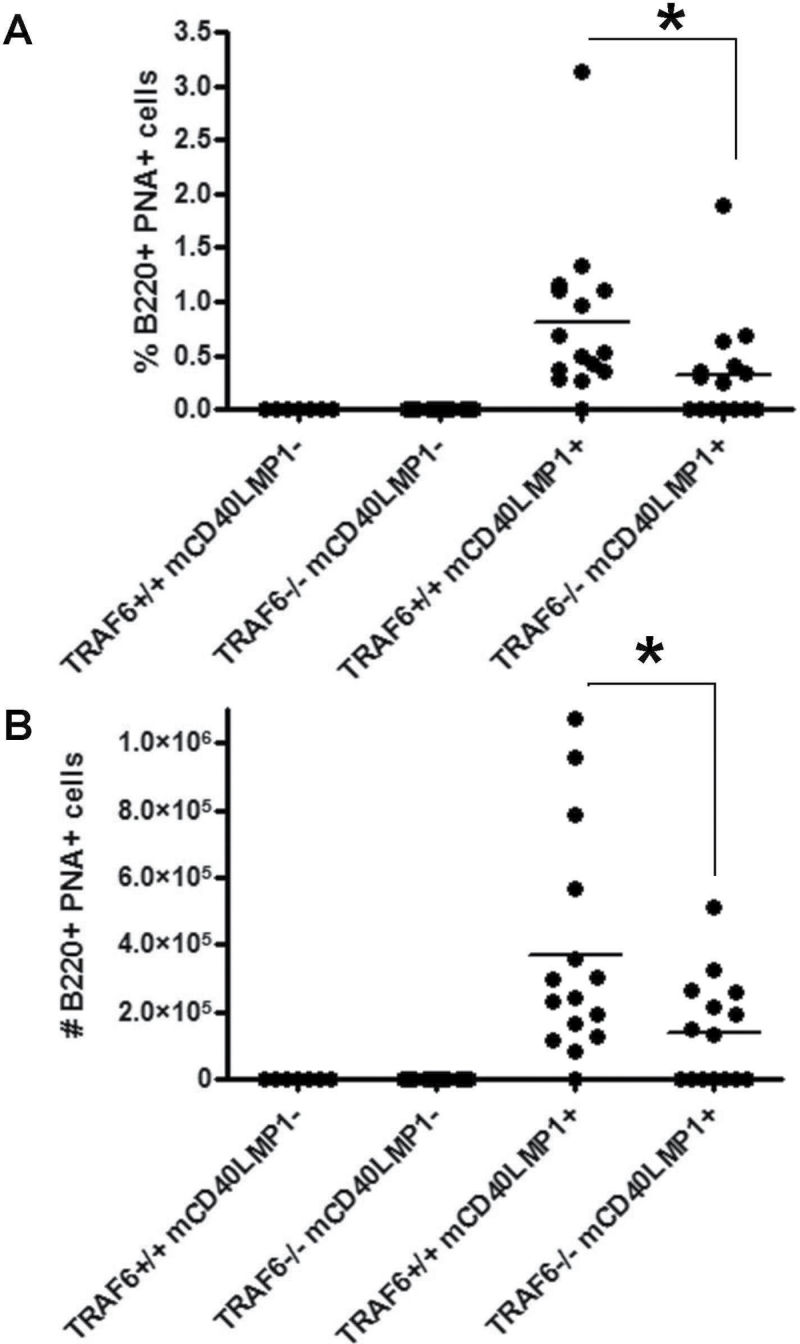
Impact of TRAF6 on spontaneous GC formation in mCD40LMP1 Tg mice. (A) Splenocytes from TRAF6+/+ mCD40LMP1-negative, TRAF6−/− mCD40LMP1-negative, TRAF6+/+ mCD40LMP1 Tg and TRAF6−/− mCD40LMP1 Tg mice were stained with anti-B220 antibody and the GC marker PNA. The percentage of B220+ PNA+ GC B cells is shown, where each point on the graph represents one mouse. (B) Similar to A, except the total number of B220+ PNA+ GC B cells is shown. *Statistical difference between TRAF6+/+ and TRAF6−/− mCD40LMP1 Tg mice as determined by the Student’s unpaired t-test, P < 0.05.

