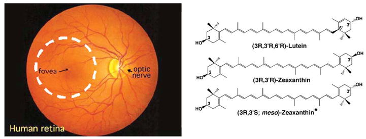
Fig. 1.

Ophthalmoscopic view of a human retina (left) showing the boundaries of the human macula as a 5 mm diameter dashed white circle centered on the fovea. The macular carotenoid pigment is concentrated in the central 500 microns of the macula at the fovea. The chemical structures of the major macular pigment carotenoids are shown on the right.
