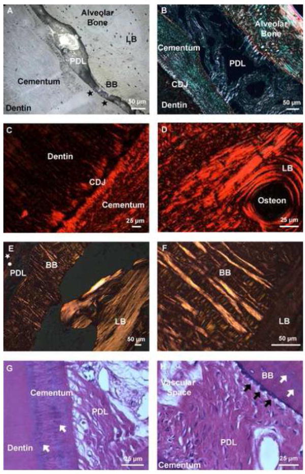
Figure 2. Fiber orientation varied across tissues of the bone-PDL-cementum complex.

Higher magnification light micrograph of an ultrasectioned surface block illustrated structural heterogeneity in alveolar bone, PDL, and tooth. The tissue-level characteristics of constricted PDL-space include invasion of BB from LB and adapted cementum (black stars) (A). Picrosirius red stain coupled with polarized light microscopy of the narrowed region demonstrated differential fiber orientation within secondary cementum and alveolar bone (B), and the differential collagen fiber orientation between the cementum dentin junction (CDJ), and lamellae and interlamellae regions (characterized by birefringence) (C, D). Lower and higher magnification hematoxylin and eosin staining (H&E) of alveolar bone coupled with polarized light microscopy demonstrated the heterogeneity of fiber orientation between BB, which was dominated by radial fibers, versus LB consisting of primarily circumferential fibers (circle: cementum, white star: dentin) (E,F). Hematoxylin and eosin staining (H&E) demonstrated localization of basophilic lines in both cementum and bundle bone (white arrows). Additionally, directional bone growth illustrated by bony protrusions (black arrows) was expressed at the PDL-bone interface (G,H). LB: Lamellar bone, BB: Bundle bone, PDL: Periodontal ligament, CDJ: Cementum-dentin junction. *Note: the orientation of the bone-PDL-tooth complex varies in images.
