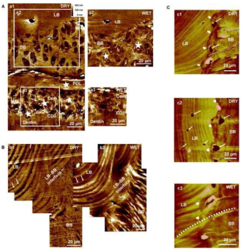
Figure 3. Tissue architecture under dry and wet conditions illustrated LB-BB interface as the original site of PDL attachment.

Higher resolution AFM micrograph of an alveolar bone-PDL-cementum complex imaged under dry (A: a1) and wet (A: a2, a3) conditions. Radial fiber insertions (stars, cross sections revealed) into both BB and cementum interfacing the PDL are visible. Swelling of insertion fibers (stars) under wet conditions are indicative of polyanionic molecules and predominance of organic matter. The LB-BB interface is shown in greater detail under dry (B: b1) and wet (B: b2) conditions. Under dry conditions, lamellae regions demonstrated greater depth relative to inter-lamellae regions of LB. However, under wet conditions lamellae regions demonstrated increased hygroscopicity compared to inter-lamellae regions of LB, suggestive of differential degrees of mineral and/or organic content. A corresponding lamellar region is denoted by arrows in both dry and wet conditions. It should be a noted that angle of sectioning can illustrate a structure significantly different from the LB-BB interface, as observed in B: b2. Additionally, continuous fibers from BB through the distinct interface and attached to LB are visible (C: c1, c2, c3). All images exhibited hygroscopicity under wet conditions (C: c3) and insertion of Sharpey’s fibers across BB and into the LB region, alongside multiple osteocytes, indicates the LB–BB interface as the point of genesis for BB growth. LB: Lamellar bone, BB: Bundle bone, PDL: Periodontal ligament, Cem: Cementum, CDJ: Cementum-Dentin Junction.
