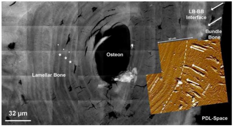
Figure 5. A higher resolution map of heterogeneous X-ray attenuations within alveolar bone.

A composite made out of micrographs taken using a high resolution Nano-TXM illustrated varying X-ray attenuations within bundle bone (BB). Bone adjacent to the PDL-space was identified as BB by the presence of less attenuating organic Sharpey’s fibers inserts (white arrows). Interestingly, the LB-BB interface is highlighted as highly X-ray attenuating suggesting higher mineral content. Overlay is an AFM micrograph with radial PDL fibers in BB and an orientation change to circumferential in LB within a junction of 10-30 μm. It is possible that the density of fibers could have contributed to the higher X-ray attenuation (dashed white line). Highly attenuated (radiopaque) regions (star) in lamellar bone (circle). Lamellar bone: LB, Bundle bone: BB, Periodontal ligament: PDL
