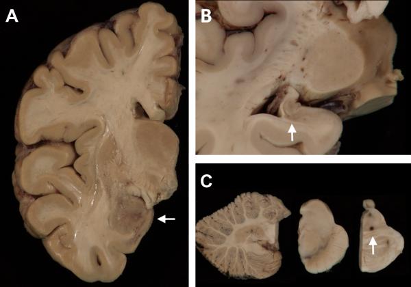
FIGURE 1.
Macroscopy. (A) A coronal slice of the right hemibrain shows discoloration and granularity of the amygdala (arrow). A photomicrograph of the left hippocampus (B) shows diminished size and gray discoloration (arrow). (C) The cerebellum is unremarkable (left panel) and the substantia nigra (center panel) and locus coeruleus of the pons (right panel) show no significant pallor. There is an area of dark-brown discoloration at the junction of the tegmentum and basis pontis which corresponded to a nidus of bacteria-containing macrophages seen microscopically.

