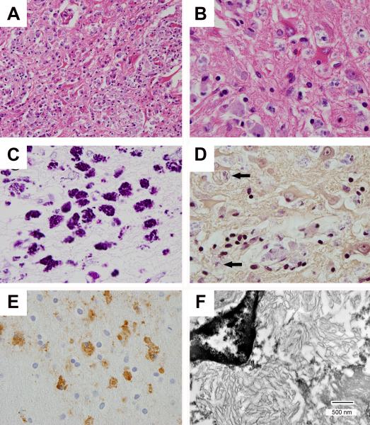
FIGURE 2.
Microscopy and fine structure of T. whipplei encephalitis. There is parenchymal disruption, reactive astrocytosis, and macrophages with basophilic staining of intracellular material (A, hematoxylin and eosin (HE) × 400; B, HE × 1,000), and a granular staining pattern with periodic acid-Schiff (PAS) stain (C, × 1000). Gram stain shows predominantly pale blue staining of intracellular debris with occasional foci of strong gram-positivity (D, arrows: Gram stain × 1,000). Anti-T. whipplei antibodies highlight macrophages (E, T. whipplei immunohistochemistry × 1,000), and transmission electron microscopy reveals lamellar structures consistent with degraded bacteria (F, bar = 500 nm).

