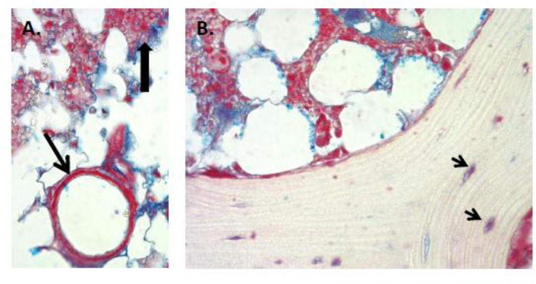
Figure 5. Perlecan staining in human bone, bone marrow and basement membrane of bone marrow endothelial cells.
A. Perlecan staining (red) is clearly seen in bone marrow, particularly in some cells (dark arrowhead), and intense in the basement membrane surrounding bone marrow capillaries (arrow). Blue is GAG staining with Alcian Blue. Any cells entering or leaving the circulation would need to traverse the perlecan rich border underlying the capillary endothelial cells. B. Perlecan staining is seen at the endosteal border, surrounding osteocytes in dense bone (arrows), and in the reticular network supporting hematopoiesis in bone marrow, but not in mineralized bone. (Images courtesy George Dodge).

