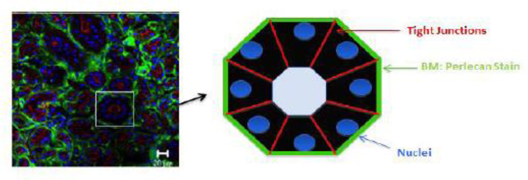
Figure 6. Perlecan expression in the human salivary gland basement membrane.
Primary tissue from a freshly excised patient specimen was stained with an antibody recognizing perlecan (green) or the tight junction marker, ZO-1 (red). The nuclei are shown in blue. The cartoon illustrates the concept that perlecan in the basement membrane separates and surrounds each structural unit, separating structures from stroma and each other, forming discrete borders around each secretory unit. (Figure courtesy Dr. Swati Pradhan-Bhatt).

