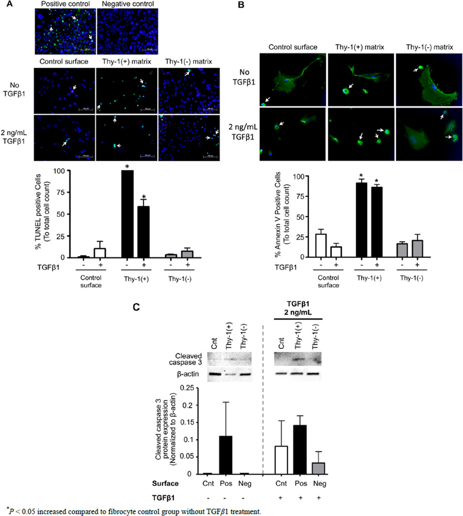
Figure 7.
The extracellular matrix (ECM) derived from Thy-1(+) positive lung fibroblasts promotes apoptosis pathways in fibrocytes. Cultured fibrocytes were harvested and analyzed for markers of apoptosis by (A) TUNEL assay, (B) Annexin V expression, and (C) cleaved Caspase 3 protein expression 72 hours after TGFβ1 treatment. The upper inset in (A) shows representative microscopic images of the TUNEL assay(200×, bar = 100 µM). The lower inset in (A) is a bar graph representation of the percentages of TUNEL positive cells from 5 random 200× magnification fields per slide. The upper inset in (B) shows representative microscopic images of Annexin V staining (200×, bar = 100 µM) and the lower inset is a bar graph representation of the percentages of Annexin V positive cells analyzed from 6 random 200x magnification fields per slide. White arrows = cells positive for Annexin V. The upper inset in (C) shows Caspase 3 protein expression by western blot and the lower inset is a bar graph showing relative Caspase 3 protein expression by densitometric analyses. Cnt = Control, Pos = matrix derived from Thy-1(+) fibroblasts, and Neg = matrix derived from Thy-1(−) fibroblasts. N = 3 mice per group. Data are represented as mean ± SEM.

