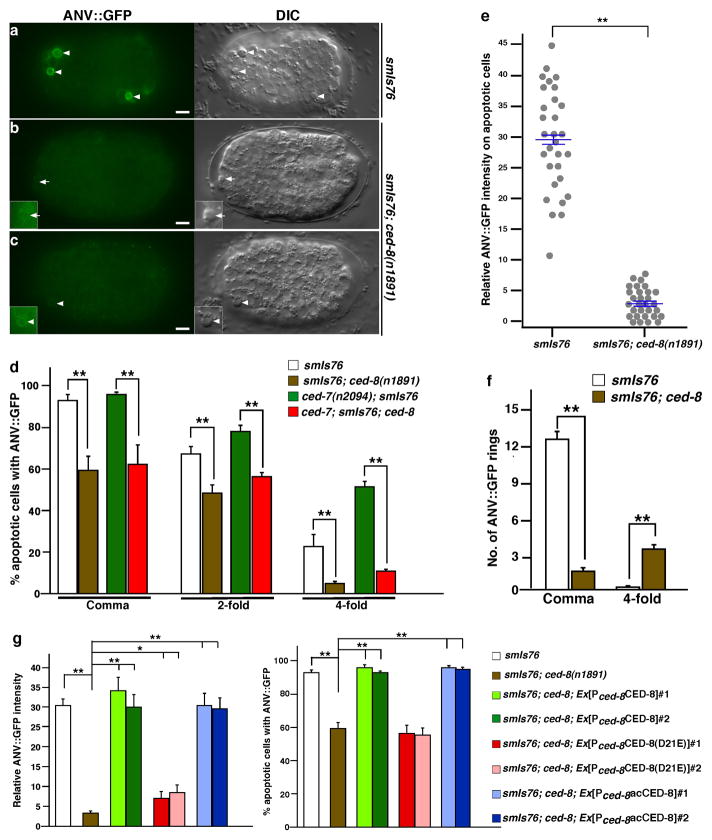
Figure 2. PS externalization in apoptotic cells is blocked or greatly reduced in the ced-8 mutant.
(a–c) ANV::GFP and DIC images of comma stage embryos from smIs76 and smIs76; ced-8(n1891) animals are shown. Arrowheads indicate apoptotic cells labeled by ANV::GFP. In smIs76; ced-8(n1891) embryos, apoptotic cells either were not labeled (indicated by an arrow, b) or were very weakly labeled (c) by ANV::GFP. Insets show enhanced GFP images of apoptotic cells. Scale bars indicate 5 μm. (d) Percentages of apoptotic cells labeled by ANV::GFP in the indicated strains and stages are shown. (e) Relative intensity of ANV::GFP that labeled apoptotic cells in comma stage embryos was quantified in the indicated strains (see Methods). Results are shown using the histogram (n=30), with mean ± s.e.m. indicated. Apoptotic cells that were not labeled by ANV::GFP are presented with zero intensity. (f) The number of ANV::GFP rings in the indicated strains was scored in the comma and 4-fold stage embryos regardless of their cell morphology. (g) Rescue of the PS externalization defect of the ced-8(n1891) mutant by various ced-8 transgenes. Quantification of the percentage of apoptotic cells labeled by ANV::GFP and the relative intensity of ANV::GFP in transgenic embryos was performed as in (d, e). (d, f, g) Results are presented as mean ± s.e.m. (n=15 each). The significance of differences between results was determined by two-tailed Student’s t-tests, **P < 0.001; *P < 0.05. Heat-shock treatment was performed at 33°C for 30 min and embryos were examined after 2 hour recovery at 20°C. Exposure time for each fluorescent image was identical (12.5 ms).

