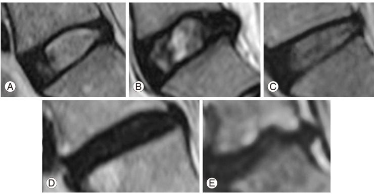
Fig. 4.
Pfirrmann's grading of disc degeneration on T2-weighted magnetic resonance imaging (MRI) image. (A) Grade 1: nucleus appears homogenously white, disc height is preserved, clear distinction between nucleus and annulus. (B) Grade 2: similar to grade 1 except for some signal intensity changes in the nucleus like horizontal clefts. (C) Grade 3: nucleus appears gray, disc height preserved or slightly reduced, distinction between nucleus and annulus is unclear. (D) Grade 4: nucleus appears black, with moderate reduction in the disc height without any distinction between nucleus and annulus. (E) Grade 5: completely collapsed homogenously black disc.

