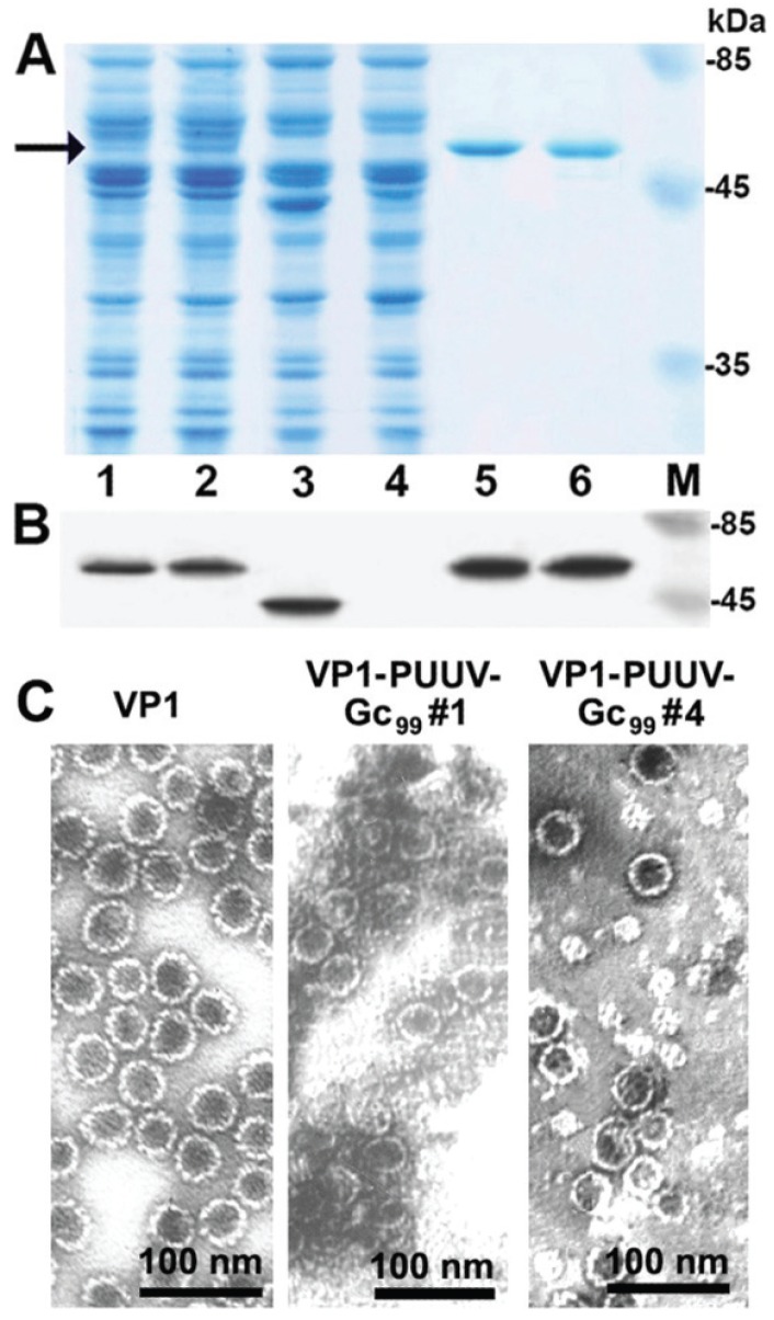
Figure 3.
Analysis of VP1-PUUV-Gc99 fusion proteins by SDS-PAGE (A) and Western blot using VP1-specific MAb 6D11 (B). Lane 1, crude lysate of yeast cells transformed with pFX7-VP1/L/Kaz-Gc99#1 plasmid; lane 2, crude lysate of yeast cells transformed with pFX7-VP1/L/Kaz-Gc99#4 plasmid; lane 3, crude lysate of yeast cells transformed with pFX7-VP1 plasmid; lane 4, negative control sample from crude lysate of S. cerevisiae cells transformed with “mock” vector pFX7; lane 5, VP1-PUUV-Gc99#1 VLPs after purification in sucrose and CsCl gradients; lane 6, VP1-PUUV-Gc99#4 VLPs after purification in sucrose and CsCl gradients; lane M, prestained protein weight marker (UAB “Thermo Fisher Scientific Baltics”); (C) Electron microscopy pictures of VP1 and VP1-PUUV-Gc99 VLPs stained with 2% aqueous uranyl acetate solution and examined by a JEM-100S electron microscope.

