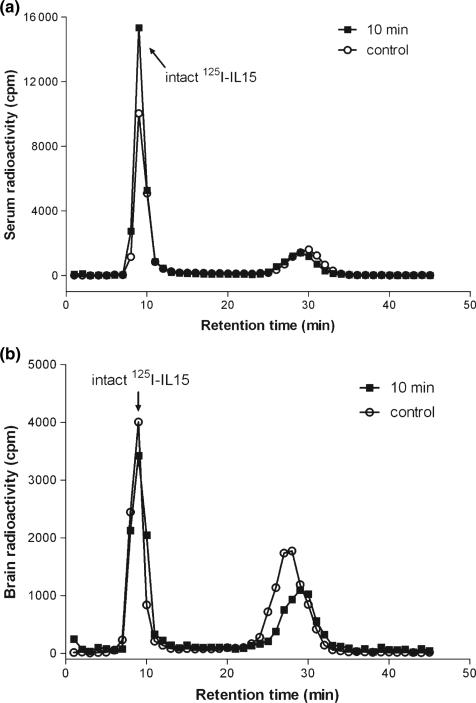
Fig. 1.
(a) Size-exclusion chromatography of serum radioactivity 10 min after i.v. injection of 125I-IL15. The control involved collection of blood into a test tube containing the 125I-IL15 to assess the extent of ex vivo degradation. In both samples, intact 125I-IL15 represented more than 95% of the total radioactivity, the rest being free 125I. (b) Size-exclusion chromatography of radioactivity in brain homogenate 10 min after i.v. injection of 125I-IL15. The control involved homogenization of an untreated brain in a homogenizer containing 125I-IL15. Intact 125I-IL15 accounted for 95% of the total radioactivity in these samples. IL, interleukin.

