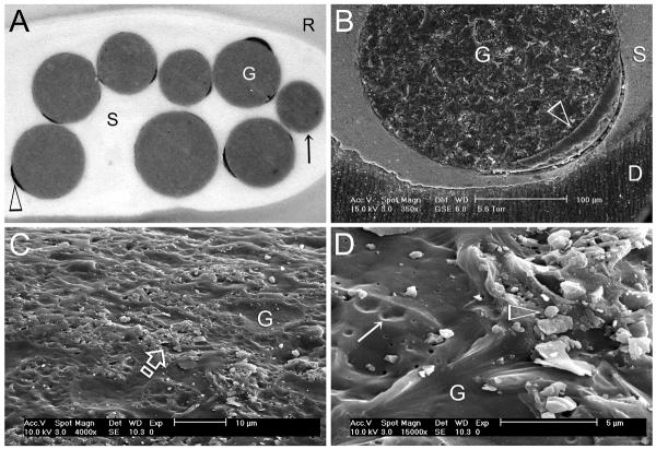
Figure 1.
A. Stereoscopic microscope image of incomplete coating (open arrowhead) of resin-coated gutta-percha points (G) that are used with a hydrophilic self-priming methacrylate resin-based root canal sealer (S). Some obturation points are devoid of resin coating (arrow). B. Environmental scanning electron microscopy (ESEM) image of a partially resin-coated gutta-percha point (G), taken at 95% relative humidity, showing a gap between the resin coating (open arrowhead) and sealer; D: root dentin. C. SEM of the surface of a bioactive glass particle-coated gutta-percha point (G) that is used with a glass ionomer-based root canal sealer. Distribution of the glass particles is limited to some areas only (open arrow). D. High magnification of Figure 1C. Area on the left is devoid of glass particles (open arrowhead). Areas previously occupied by glass particles (open arrowhead) are denoted by depressions on the gutta-percha surface (arrow).

