
Figure 2.
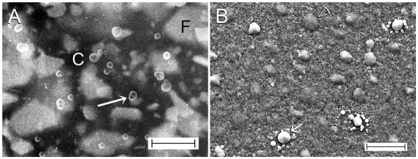
A. SEM image of the heat-pressed surface of thermoplastic polycaprolactone-based root filling material (C) showing phase separation of the methacrylate resin component (arrow) from the polycaprolactone. F: fillers. Bar = 5 μm. B. SEM image of the polycaprolactone-based root filling material after surface etching of the polycaprolactone root filling material with 0.1 N NaOH revealing exposed, partially-coalesced methacrylate resin droplets (arrow). Open arrowhead: unexposed fillers. Bar = 20 μm.