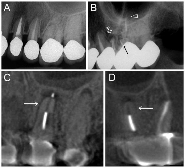
Figure 4.
A. Pre-operative periapical radiograph of the upper left first molar (second tooth from the right). Although the tooth was non-vital, there was no periapical radiolucency associated with the palatal root. B. Post-operative periapical radiograph of the upper left first molar after root canal treatment and obturation with a polycaprolactone-based root filling material and accompanied resin-based sealer. Four radiopaque filled canals can be identified: 2 canals in the mesiobuccal root (open arrow), one canal in the distobuccal root (arrow) and one canal in the palatal root (open arrowhead). C. Three-year post-treatment cone beam computer tomography image (sagittal projection across the palatal root) of the same root-treated upper left first molar showing partial disappearance of the polycaprolactone root filling material (arrow) and the development of a periapical lesion at the root tip of the palatal root. D. Three-year post-treatment cone beam computer tomography image of the same tooth taken along an oblique coronal projection, revealing the palatal root and the distobuccal root. Disappearance of the polycaprolactone root filling material from the apical third of the root canal (arrow) is also evident from this angulation.

