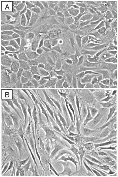
Fig. 2.
Morphology of human articular chondrocytes grown in monolayer culture on plastic. Chondrocytes were isolated from articular cartilage and cultured in DMEM/F12 containing 10% FCS until confluent and then subcultured and grown again to confluence. (a) Primary chondrocytes display the characteristic cobblestone morphology. (b) Passaged chondrocytes display features of a dedifferentiated phenotype, including a portion of cells with fibroblast-like morphology.

