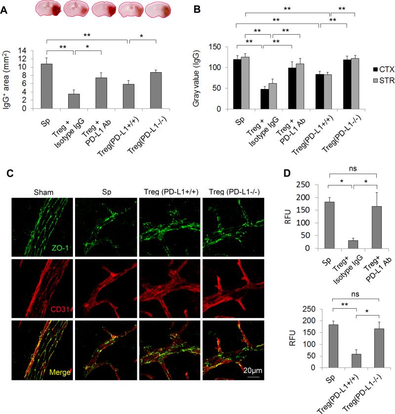
Figure 5. PD-L1 is critical for Treg-afforded BBB preservation after MCAO.
To block PD-L1 signaling in Tregs, Tregs were pre-incubated with PD-L1 neutralizing Abs or prepared from PD-L1 knockout mice and then adoptively transferred into MCAO animals after 2 h of reperfusion. Isotype IgG-treated or wild-type Tregs were used as controls, respectively. (A-B) Quantification of IgG extravasation at 24 h after MCAO. (A) Quantification of endogenous IgG positive area determined by immunohistochemical staining of mouse IgG. n=5/group. (B) Quantification of gray values of IgG immunostaining in the cortex (CTX) and striatum (STR). n=5/group. (C) Representative Z-stack confocal images of the tight junction protein ZO-1 and endothelial cell marker CD31 in brain sections obtained 1 day after MCAO. Images are representative of brain sections from four mice in each group. (D) Cadaverine-Alexa-555 (950Da) was injected intravenously 22 h post-MCAO. Fluorescence intensities in brain lysates from the infarct area were measured after 2 h of circulation. n=5/group. *P<0.05, **P<0.01. Sp = splenocytes.

