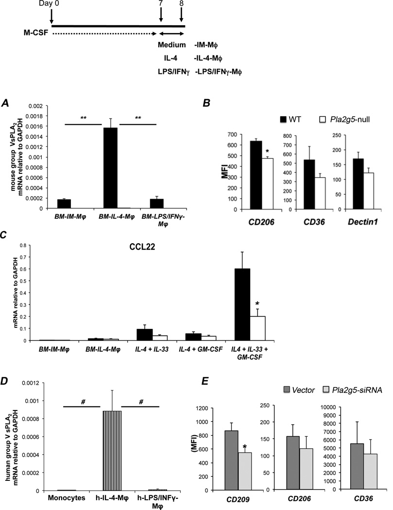
Figure 4. Polarization of Mϕ toward alternative activation requires gV-sPLA2 in mouse and human.
(A) Expression of gV-sPLA2 mRNA relative to GAPDH measured by qPCR in WT (filled bars) and Pla2g5-null (open bars) mouse BM-IM-Mϕ or BM-IM-Mϕ polarized with IL-4 or LPS/IFN-γ (BM-IL-4-Mϕ and BM-LPS/IFNγ-Mϕ, respectively). (B) Net (isotype control subtracted) MFI of CD206, CD36, and Dectin1 on CD11b+/F480+ WT (filled bars) and Pla2g5-null (open bars) mouse BM-IL-4-Mϕ evaluated by flow cytometry. (C) Expression of CCL22 relative to GAPDH in BM-IM-Mϕ, BM-IM-Mϕ polarized with IL-4, IL-4+IL33, IL-4+GM-CSF and IL-4+IL-33+GM-CSF measured by qPCR. (D) Expression of human gV-sPLA2 mRNA relative to GAPDH in monocytes, IL-4 and LPS/IFN-γ polarized human monocyte-derived Mϕ (h-IL-4-Mϕ and h-LPS/IFNγ-Mϕ, respectively) measured by qPCR. (E) Transfection of h-IL-4-Mϕ with human Pla2g5-siRNA (light gray columns) or non-targeting vector (dark gray columns) (1000 nM) and net (isotype control subtracted) MFI of CD209, CD206, and CD36 on gated CD11b+ cells evaluated by flow cytometry. Values are mean ± SEM of three (A, C, D and E), or four to six (B) independent experiments. *, p<0.05; #, p<0.002; **, p<0.001

