Fig. 6.
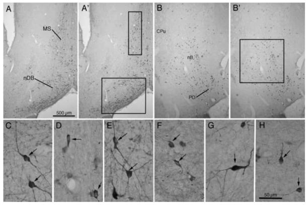
ChAT immunoreactivity in basal forebrain. Photomicrographs in A and B show patterns of immunoreactivity, while A′ and B′ indicate sites of cell counts in medial septum (MS – box 300 μm × 1000 μm), nucleus of the diagonal band (nDB – box 800 μm × 1000 μm), and nucleus basalis (nB – box 1000 μm × 1000 μm). Photomicrographs in the lower row (C–H) present higher magnification images of ChAT positive cells in these basal forebrain regions. Normal appearing cells (type 1) from animals treated with saporin followed by NT3 are shown in C (MS), E (nDB), and G (nB). Cells appearing atrophic (type 2) from animals treated with saporin followed by saline are shown in D (MS), F (nDB), and H (nB). Scale bar in A = 500 μm for A, A′, B, and B′. Scale bar in H = 50 μm for C – H.

