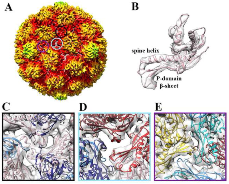
Figure 1. Subnanometer CryoEM reconstruction of protease-free HK97 Prohead-1.

(A) Surface rendering of the procapsid reconstruction low-pass filtered at 7.8 Å resolution, sharpened with a B factor of -700 Å2 and radially colored as in Figure 1. The three circles overlaid onto the particle correspond to the three classes of 3-fold contacts and their respective color point to the corresponding frame in panels (C), (D) and (E). The reconstruction is radially colored according to the color key used in Figure S1. (B) Fit of a Prohead-1 coat subunit (PDB ID 3QPR) into the corresponding region of the reconstruction shown in (A). The seven subunits of the icosahedral asymmetric unit are conformationally distorted at the level of the spine helix and P-domain β-sheet. (C) Class I icosahedral 3-fold contacts involving subunits D. (D) Class II quasi-3-fold contacts involving subunits B, C and E. The coat subunit N-arms have been removed for clarity on this panel. (E) Class III quasi-3-fold contacts involving subunits A, F and G. The coat subunit coordinates are colored as follows: A (cyan), B (dark red), C (red), D (pink), E (navy blue), F (dodger blue) and G (gold). Panels C-E represent views from the virion interior.
