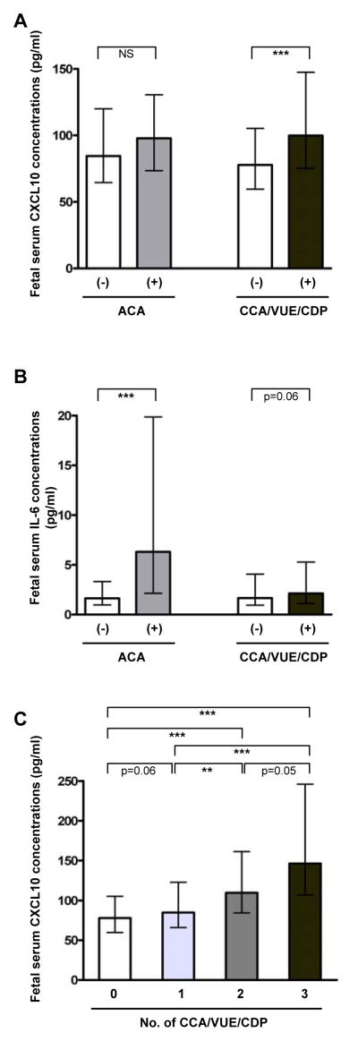
Figure 1. Fetal serum CXCL10 and IL-6 concentrations according to the presence or absence of maternal anti-fetal cellular rejection.
(A) Fetal serum CXCL10 concentration was higher in cases with anti-fetal cellular rejection (chronic placental inflammation) than in those without (P<0.001), while fetal serum CXCL10 concentration was not different according to the presence or absence of acute chorioamnionitis. (B) Cases with acute chorioamnionitis had higher median fetal serum IL-6 concentration than those without (P<0.001), and fetal serum IL-6 concentration tended to be higher in cases with anti-fetal cellular rejection than in those without (P=0.06). (C) The upward trend of blood CXCL10 concentration correlates with the extent of cellular rejection (P<0.001 by the Jonckheere-Terpstra test). Fetal serum CXCL10 and IL-6 concentrations were shown as median and inter-quartile ranges. *P<0.05; **P<0.01; ***P<0.001 (by the Mann-Whitney U test for comparison between the two groups). ACA, acute chorioamnionitis; CCA, chronic chorioamnionitis; CDP, chronic deciduitis with plasma cells; NS, not significant; VUE, villitis of unknown etiology.

