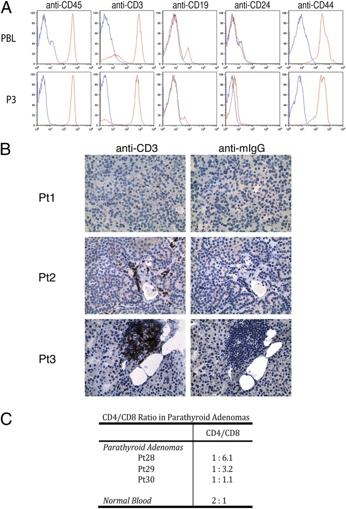
Fig. 3.
Immunophenotypic characterization of cells isolated from parathyroid adenomas. (A) Flow-cytometric analysis of cell-surface markers CD45, CD3, CD19, CD24, and CD44 on tumor-infiltrating lymphocytes (TILs) (Lower) and patient-matched PBLs (Upper). (B) Tissue sections from three independent parathyroid adenomas were probed with an anti-CD3 antibody, and reactivity was visualized by DAB staining and with hematoxylin/eosin counterstaining. (C) CD4 and CD8 cell-surface expression in P3 cells derived from three parathyroid adenomas was determined by FACS. The ratio of CD4+ to CD8+ cells is based upon quadrant gating using standard conditions.

