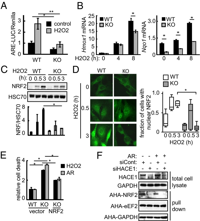
Fig. 2.
HACE1 promotes NRF2 activity, stability, and synthesis. (A) Hace1 WT or KO MEFs cotransfected with antioxidative stress response element-luciferase (ARE-LUC) and pRenilla vectors were treated with H2O2 (200 μM; 6 h), as indicated. NRF2 activity was determined by measuring luciferase activity. n = 3; *P < 0.01; **P < 0.005. (B) Hace1 WT or KO MEFs were treated with H2O2 (200 μM) for the indicated times. Hmox1 and Nqo1 mRNA levels were determined using qRT-PCR. n = 3; *P < 0.01. (C) Hace1 WT or KO MEFs were treated with H2O2 (200 μM) for the indicated times. NRF2 protein levels were determined using Western blot. NRF2 levels were normalized to HSC70. n = 3; *P < 0.05. (D) Hace1 WT or KO MEFs were treated with H2O2 (200 μM) for the indicated time points. NRF2 localization was determined using immunofluorescence. Typical images are shown on the left. Cells exhibiting nuclear or cytosolic localization were scored. n = 200 from three independent experiments; *P < 0.01. (E) Hace1 WT or KO MEFs transfected with NRF2-expressing or control vector were treated with H2O2 (600 μM) or AR (10 μM) for 8 h. Cell death was measured by annexin V and PI staining and presented relative to WT cells. n = 3; *P < 0.01. (F) HEK293T cells were transfected as in E. Cells were pulsed with AHA (250 μM; 1 h) and MG132 to block protein degradation with or without AR (1 μM) as indicated. HACE1 kd in total cell lysates was confirmed using Western blot. Cell lysates were subjected to a Click-it reaction with biotin alkyne followed by streptavidin pulldown, and newly synthetized proteins were detected by Western blot.

