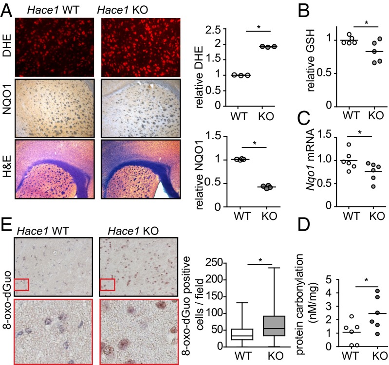
Fig. 3.
HACE1 loss is associated with reduced antioxidants and increased oxidative stress in brain. (A) The striatal regions of Hace1 WT and KO brains were stained using dihydroethidium (DHE), anti-NQO1 antibodies, or H&E as indicated. Sections were imaged using a fluorescent (DHE) or light microscope (NQO1 and H&E). Staining was quantified using ImageJ. n = 3; *P < 0.05. (B) GSH levels were measured in brain lysates from Hace1 WT and KO mice. Values were normalized to WT. *P < 0.05. (C) Nqo1 mRNA levels in mouse brains determined by qRT-PCR. *P < 0.01. (D) Protein carbonylation was measured in Hace1 WT and KO mouse brains. *P < 0.05. (E) Hace1 WT and KO mouse brains were subjected to anti–8-oxo-dGuo immunohistochemistry. Staining in the cortex was quantified using ImageJ. *P < 0.001.

