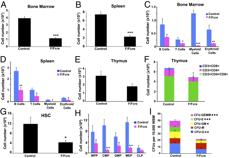
Fig. 2.
B-myb is required for hematopoiesis. B-myb–deficient (F/Fcre; Mx1-cre-B-mybF/F) and control (B-myb+/F or B-mybF/F) mice were treated with pIpC every other day over a 5-d period. Tissues were harvested on day 21 posttreatment. Total number of cells isolated from BM (A) and spleen (B) of control and B-myb–deficient mice. Analysis of mature lineage cells present in the BM (C) and spleen (D) of control and B-myb–deficient mice. Erythroid, B-, T-, and myeloid lineage cells are defined as TER119+, B220+, CD3+, and CD11b+Gr1+, respectively. Total number of thymocytes (E) and mature populations (F) in the thymuses of control and B-myb F/Fcre mice. (G) Total number of HSCs in the BM of control and B-myb–deficient mice. (H) Total number of MPPs, myeloid progenitors (CMPs, GMPs, and MEPs), and lymphoid progenitors (CLPs) in the BM of control and B-myb–deficient mice. (I) Colony-forming assay showing the frequency of whole BM cells that formed megakaryocyte (GEMM), erythroid (E), granulocyte–monocyte (GM), monocyte (M), or granulocyte (G) colonies in culture. All values represent mean ± SEM. n ≥ 3 per population for each genotype. *P ≤ 0.05, **P ≤ 0.005, ***P ≤ 0.0005.

