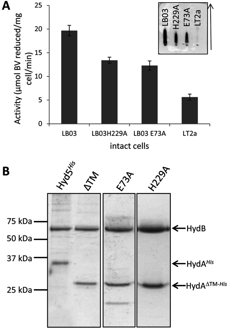
Figure 2. Isolation of S. enterica Hyd-5 and its variants.
(A) The S. enterica strains LT2a (parent strain), LB03 (PT5, hydAΔTM−His), LB03 H229A and LB03 E73A were grown anaerobically and whole cells were assayed for hydrogen-dependent BV reduction activity. Results are means±S.E.M. (n=3). Inset, anti-Hyd-5 rocket immunoelectrophoresis of periplasmic fractions prepared from the strains LB03 (PT5, hydAΔTM−His), LB03 H229A, LB03 E73A and the parental strain LT2a. The arrow shows the direction of protein migration. (B) SDS/PAGE (12% w/v gel) of pooled IMAC fractions of proteins isolated from strains SFTH06 (PT5, hydAHis), LB03 (PT5, hydAΔTM−His) (‘ΔTM’), LB03 H229A and LB03 E73A. Images taken from different gels are separated by black borders. Molecular mass is given on the left-hand side in kDa.

